In medical imaging, maintenance of image quality at clinically acceptable levels to allow for accurate disease diagnosis is important. Image quality can be evaluated with an imaging phantom, i.e. a specially designed object that can be imaged by medical imaging devices such as x-ray machines or computed tomography (CT) scanners. Imaging the phantom allows evaluators to evaluate, analyse and optimise the performance of the imaging devices to maintain clinically relevant image quality levels. This process should be done regularly to keep devices working at their best. The phantom is used instead of living subjects because the phantom gives more consistent results and avoids unnecessarily exposing patients to excess radiation.
Phantoms provide a consistent method to monitor quality of imaging devices in terms of contrast, resolution and noise of the outputs. However, currently each type of image device has dedicated phantoms and associated computer software commercially available. This is costly and complicated to maintain, especially for resource limited medical institutions where cost, manpower, expertise and time constrains are identified problems. The U-QA universal radiology x-ray phantom can be used in a variety of imaging devices to provide a simpler and more cost-effective solution.
The U-QA phantom will solve the problem of having to purchase, learn and maintain different proprietary phantoms and associated software for each imaging device. The U-QA phantom, associated software and comprehensive manual will allow users to evaluate and optimize the performance of their imaging devices to ensure optimal results and the best care for patients. Specifically the phantom will allow qualitative and quantitative tests of sensitometry, uniformity, resolution, noise, geometry, standard signal, low contrast detectability, alignment, artefacts, visual image quality inspection, CT slice thickness and mammography masses, fibres and micro-calcification detection. It is applicable in general x-rays, fluoroscopy, mammography, and CT scanning. The phantom will assess the image quality parameters stipulated by the Department of Health, Directorate Radiation Control, in diagnostic radiology equipment licenses.
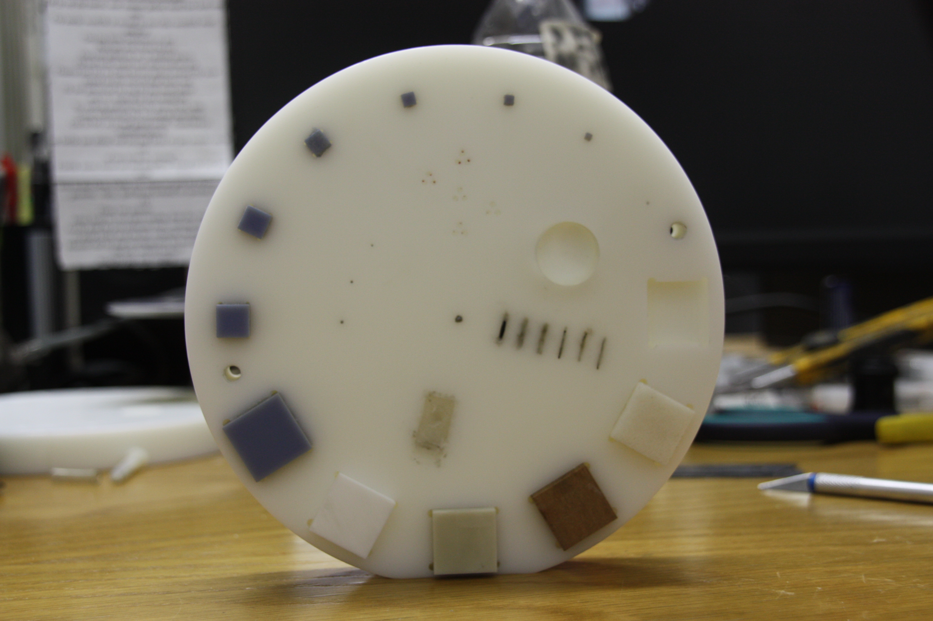
The phantom can be set up vertically for CT scanning or horizontally for planar imaging, i.e. general x-rays, mammography and fluoroscopy. The phantom is manufactured from hard, radio-translucent plastic. It contains layers of inserts which are used to quantify image quality. Blocks of different density materials are used to evaluate sensitometry. For low contrast detectability, blocks of the same material but different sizes are used. Scribe lines on the phantom allows laser and cross-wire checks. A central placed bead is used to evaluate set-up and CT zero-slice position. Resolution is evaluated with modulation transfer functions (MTF) calculations from beads. Signal-to-noise, contrast-to-noise ratios and image uniformity is calculated with region of interest analysis. This is all described in a data analysis computer program, and instructions are provided in a comprehensive user manual.
Diagnostic radiology divisions in private and state hospitals and institutions
Any institution using x-ray imaging, fluoroscopy, mammography and CT scanning (focus on resource-limited institutions)
International radio diagnostics companies

Innovus, Stellenbosch University
|
15 De Beer Street
Stellenbosch
7600
South Africa
|
PO Box 3135
Matieland
7602
South Africa
|
|
t +27 (0) 21 808 3826
e info@innovus.co.za
e forms@sun.ac.za |